Introduction
Fluorescence-guided surgery (FGS) is an intraoperative imaging technique that uses fluorescent agents and optical filters to highlight target tissues in real time. It is particularly valuable for tumor delineation, helping surgeons achieve more complete resections while sparing healthy tissue.
Looking for compatible microscopes? See our guide to Carl Zeiss OPMI Pentero 800, 800 S, and 900 and their fluorescence options (e.g., BLUE 400, YELLOW 560, INFRARED 800).
How Fluorescence-Guided Surgery Works
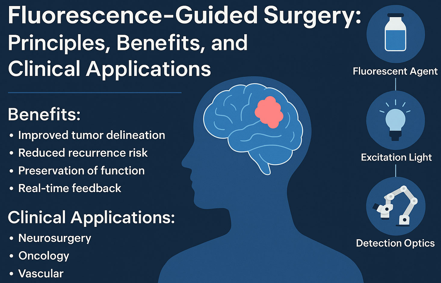
- Fluorescent agent is administered (oral, IV) and preferentially accumulates in target tissue (e.g., tumor or vasculature).
- Excitation light of a specific wavelength illuminates the field.
- Detection optics & filters capture emitted fluorescence, rendering it as a visible overlay for the surgeon.
Example: 5-ALA for Brain Tumors
Patients ingest 5-ALA, which tumor cells convert to protoporphyrin IX that fluoresces red under blue light. Surgeons visualize malignant margins in real time to guide resection.
Key Benefits
- Sharper tumor delineation vs. white-light surgery.
- Lower residual disease risk through more complete resection.
- Functional preservation by sparing eloquent, healthy tissue.
- Real-time feedback that supports intraoperative decision-making.
- Minimally invasive synergy with endoscopic, laparoscopic, and robotic workflows.
Clinical Applications
Neurosurgery
- 5-ALA (Gliolan®) for high-grade glioma visualization.
- Indocyanine Green (ICG) angiography for vessel patency and bypass assessment.
- Microscope filters such as BLUE 400 / YELLOW 560 (tumor dyes) and INFRARED 800 (NIR) on advanced systems.
Oncologic Surgery
- Margin assessment in breast, ovarian, hepatic, and colorectal procedures.
- Sentinel lymph node mapping in specific indications.
Vascular & Reconstructive
- ICG angiography for blood-flow assessment in bypass, aneurysm, flap perfusion, and organ transplantation.
For a deeper dive into operating-room workflows, see Digital Integration & PACS Workflow in the OR.
Fluorophore Comparison
| Fluorophore | Excitation (approx.) | Emission / Display | Main Clinical Use | Advantages | Limitations |
|---|---|---|---|---|---|
| 5-ALA → Protoporphyrin IX | ~405 nm (blue) | Red fluorescence | Neurosurgery (high-grade glioma) | High tumor specificity; real-time margin aid | Shallow penetration; oral dosing & photosensitivity precautions |
| Indocyanine Green (ICG) | ~805 nm (NIR) | NIR signal (often rendered green) | Vascular flow, hepatic/biliary, lymphatics | Good tissue penetration; rapid angiography; widely available | Nonspecific uptake; transient signal; iodine allergy caution |
| Fluorescein Sodium | ~490 nm (blue-green) | Yellow-green fluorescence | Ophthalmic, adjunct in tumor & vascular cases | Low cost; easy to use | Lower tumor specificity; rapid clearance; background noise |
Equipment note: If you need fluorescence-ready optics, review the Zeiss OPMI Pentero 800/800 S/900 product line.
Challenges & Limitations
- Depth limits: Most fluorescence is superficial (millimeter range).
- Agent approval: Regulatory status varies by country and indication.
- Signal variability: Inflammation or edema may cause false positives/negatives.
- System differences: Filters, sensors, and display pipelines vary by microscope/endoscope vendor.
Future Directions
- Targeted molecular probes (antibody/peptide-based) for higher specificity.
- Multispectral & ratiometric imaging to combine agents and reduce background.
- AR overlays & AI-assisted segmentation integrated into surgical displays.
Frequently Asked Questions (FAQ)
What is fluorescence-guided surgery?
FGS uses a fluorescent agent and specialized optics to highlight target tissues (e.g., tumors, vessels) in real time during surgery.
Which dyes are most common in FGS?
The most widely used agents are 5-ALA (for glioma visualization), ICG (for angiography and perfusion), and fluorescein sodium (ophthalmology and select adjunct uses).
Does FGS improve the extent of tumor resection?
FGS can improve delineation of malignant tissue, supporting more complete resections while preserving healthy structures. Outcomes depend on tumor type, surgical approach, and equipment.
Is fluorescence harmful?
The light itself is not harmful at surgical intensities. Safety considerations relate to the agents (e.g., photosensitivity after 5-ALA, iodine allergy with ICG). Surgeons follow established dosing and monitoring protocols.
Terms of Content & Medical Disclaimer
- Informational use only: This article is not a substitute for professional medical training or clinical guidelines.
- Device & agent availability: Configurations, indications, and approvals vary by region. Always verify local regulatory status and IFUs.
- No endorsement: Brand names are used for identification; inclusion does not imply endorsement.
- Clinical decisions: Must be based on the surgeon’s judgment, patient factors, and institutional policy.
- Copyright: © Your Company Name. You may quote with attribution and a do-follow link.