Used Carl Zeiss Cirrus 4000 HD OCT for sale. Excellent lightly used condition, Windows 10, sale includes the original accessories, printer, peripherals, power table and most of optional licenses has been active
This pre-owned Zeiss Cirrus 4000 fully refurbished and recalibrated by Zeiss trained technicians includes full warranty (parts and labor) and lifetime phone support.
Introducing the Zeiss CIRRUS 4000 HD-OCT, a groundbreaking addition to your practice, offering SMART OCT imaging capabilities alongside the innovative Anterior Segment Premier Module. With the latest advancements in technology, practitioners can now delve deeper into anterior segment imaging, retina analysis, and glaucoma diagnosis like never before.
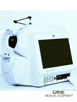
The Zeiss Cirrus 4000 HD-OCT delivers detailed diagnostic and change analysis you can rely on. The small footprint and fast capture speeds of the Cirrus 4000 improve work efficiency while helping you to deliver better care to your patients.
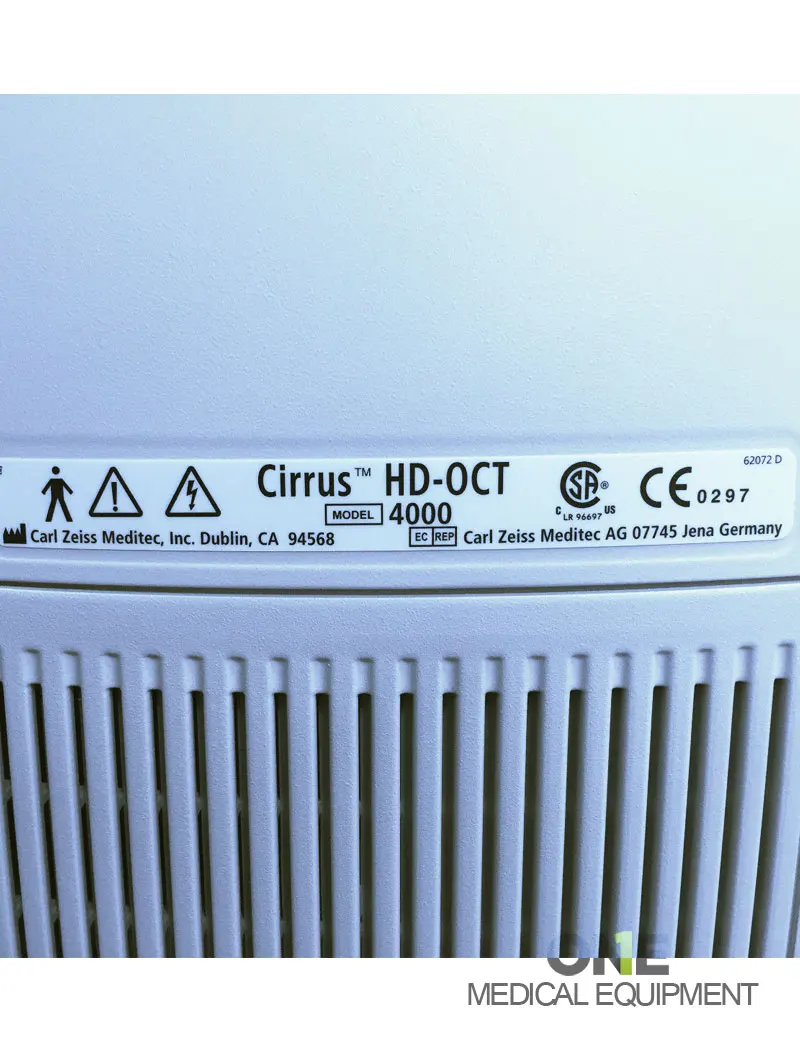
The ZEISS Cirrus HD-OCT 4000 (Cirrus HD-OCT or Cirrus) enable examination of the posterior and anterior of the eye at an extremely fine spatial scale, without surgical biopsy or even any contact with the eye. The Cirrus HD-OCT builds on and refines the retinal imaging technology first introduced with the ZEISS Stratus OCT. HD-OCT stands for ”High-definition optical coherence tomography.”
Employing the advanced imaging technology of spectral domain optical coherence tomography, Cirrus HD-OCT acquires OCT data about 70 times faster (27,000 vs. 400 A-scans per second) and with better resolution (5 μm vs. ~10 μm axial resolution in tissue), compared to first-generation OCT technology. Cirrus acquires whole cubes of OCT image data, composed of hundreds of line scans, in about the same time as Stratus acquires a six-line scan. You can view these data cubes in three planes, or through three dimensions, giving you access to an extensive amount of retinal image data in one scan.
The Cirrus HD-OCT with Retinal Nerve Fiber Layer (RNFL), Macular, Optic Nerve Head, and Ganglion Cell Normative Databases is indicated for in-vivo viewing, axial cross-sectional, and three-dimensional imaging and measurement of anterior and posterior ocular structures.
By using the advanced imaging technology of spectral domain optical coherence tomography (OCT), the Cirrus 4000 enables examination of the posterior and anterior of the eye with detailed, high-definition scans. Better assess your patient’s condition by viewing pathologies from multiple vantage points and with a range of visualization formats. The Cirrus 4000 OCT captures such a tightly packed, detail-rich cube of data in just seconds that additional scans are no longer needed.
Once a comprehensive cube of data is generated, the Cirrus can transform the information into insight. Advanced algorithms, cube tracking from visit to visit, and diversified normative databases all allow for analysis you can trust. Additionally, the Zeiss Cirrus 4000 OCT offers anterior segment imaging of the angle and cornea.
FEATURES:
The Zeiss Cirrus 4000 HD-OCT with Retinal Nerve Fiber Layer (RNFL), Macular, Optic Nerve Head, and Ganglion Cell Normative Databases is indicated for in-vivo viewing, axial cross-sectional, and three-dimensional imaging and measurement of anterior and posterior ocular structures.
The Cirrus HD-OCT is a non-contact, high-resolution tomographic and biomicroscopic imaging device. It is indicated for in-vivo viewing, axial cross-sectional, and three-dimensional imaging and measurement of anterior and posterior ocular structures, including cornea, retina, retinal nerve fiber layer, ganglion cell plus inner plexiform layer, macula, and optic nerve head.
The Cirrus normative databases are quantitative tools for comparing retinal nerve fiber layer thickness, macular thickness, ganglion cell plus inner plexiform layer thickness, and optic nerve head measurements to a database of normal subjects.
The Cirrus HD-OCT is intended for use as a diagnostic device to aid in detecting and managing ocular diseases including, but not limited to, macular holes, cystoid macular edema, diabetic retinopathy, age-related macular degeneration, and glaucoma.
Note: The Zeiss Cirrus 4000 HD-OCT is not intended to be used as the sole diagnostic for disease.
The Essential Performance of the Zeiss Cirrus 4000 is to provide accurate measurements of the anterior and posterior ocular structure.
The Cirrus HD-OCT may be used on all adults needing diagnostic eye evaluation. This includes (but is not limited to) patients with the following disabilities or challenges:
There is a general requirement that the patient sit upright and place their face in the chin and forehead for the rest of the instrument (with or without supplemental human or mechanical support).
The Zeiss Cirrus 4000 HD-OCT is designed for in-vivo viewing, axial cross-sectional imaging, and three-dimensional measurement of anterior and posterior ocular structures.
SPECIFICATIONS
Technical data
Fundus Imaging
Scan Patterns
Focus Adjustment Range
Fixation
Pupil Size Requirement
Computer
Electrical
CIRRUS 400 and 4000 systems will reach End of Support on April 30, 2022. End of Support means the end of the period where ZEISS can provide technical expertise, parts availability, and delivery of viable service to restore the product to original functionality. We will not be able to extend, renew or create any service agreements on this device. This letter is notification that all ZEISS service agreements for the CIRRUS ® 400 and 4000 systems will be terminated as of April 30, 2022. ZEISS will issue credits to customers who may have prepaid service agreements extending beyond this effective date