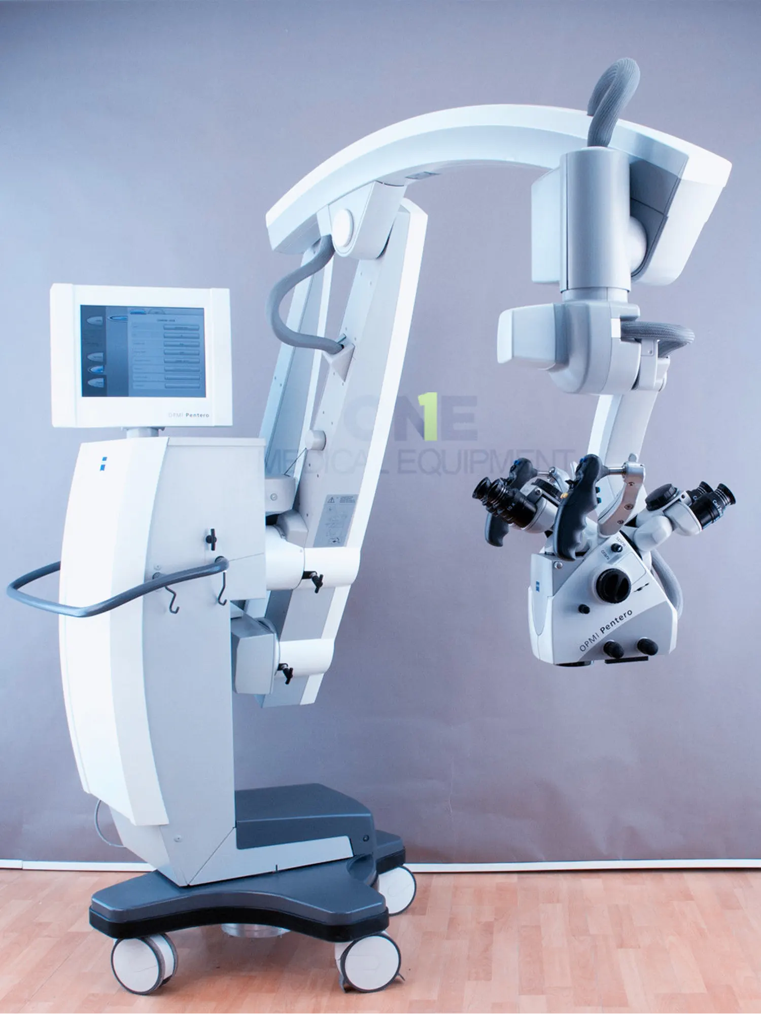
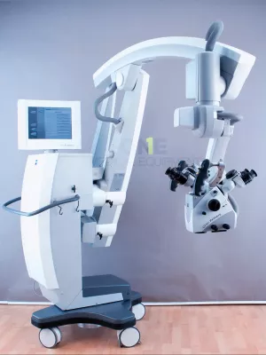
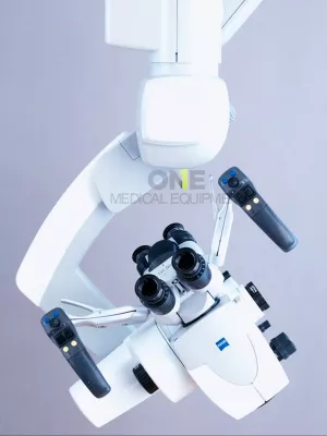
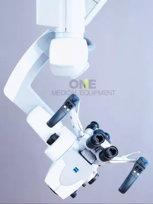
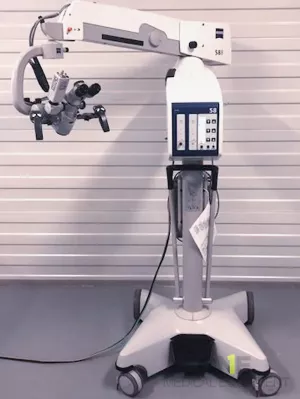
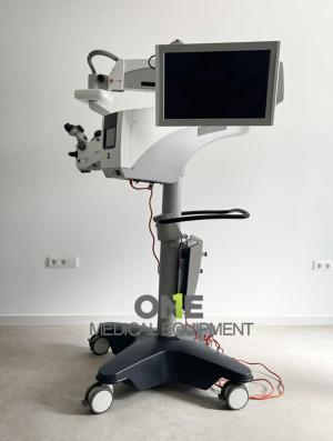
Used Carl Zeiss OPMI Pentero Floor Stand for sale, regularly serviced and maintenance by authorized Carl Zeiss service. Set up for spine, neuro and ir
Very lightly used, low hours operated at doctor office environment only, the microscope is fully functional, everything is in great and patient ready for spine, neuro and ir.
Carl Zeiss OPMI Pentero sale includes:
ACCESSORIES:
- Zeiss Blue 400
- Zeiss Infrared 800
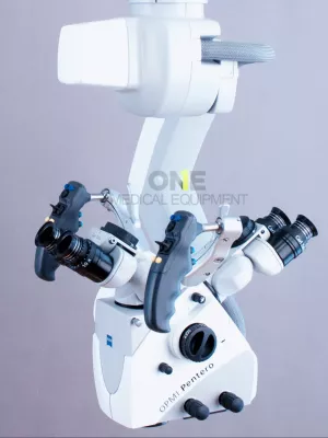
- OPMI Pentero Optics
- Stereo Bridge - Face-to-Face Bridge
- Objective lens focal length: from f=200 mm to f=500 mm (Varioskop)
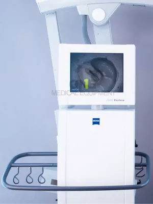
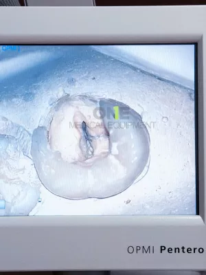
- Integrated Camera System 3CCD Zeiss Medilive
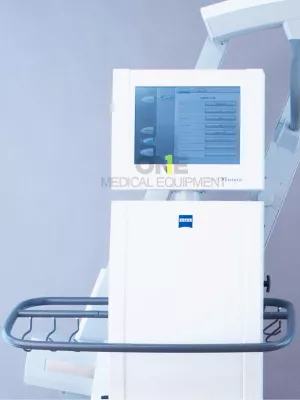
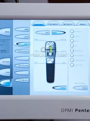
- Control unit with LCD touchscreen
- Functions:
- AutoBalance: AutoBalance of the microscope, suspension system or entire system by pushing a button
- AutoFocus: High accuracy laser guided AutoFocus that overcomes the ambiguity of the depth of field, while reliably pin-pointing the area of interest within a fraction of a millimeter
- MultiVision System: Integrated data display with shutter function
- AutoDrape System: Integrated vacuum system to remove air from sterile drape for fast and easy draping
- Back-up lamp module with Xenon lamp
- Floor Stand
- Touchscreen
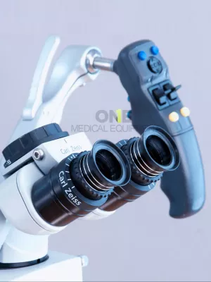
- Handgrips
TECHNICAL SPECIFICATIONS:
- Smooth zoom adjustment
- Smooth focus adjustment
- Smooth adjustment of zoom and focus displacement speed
- Smooth movement of microscope arms and microscope head (magnetic clutches)
- Smooth light adjustment
Parameters:
- Zoom adjustment:
- f=200mm: from 3,4x to 20,4x
- f=500mm: from 1,36x to 8,16x
- Magnification of the eyepieces:
- Doctor: 10x
- Assist: 10x
- Inclinable binocular tube 0°- 180° (Doctor)
- Inclinable binocular tube 0°- 180° (Assist)
- Objective lens: f=200 mm to f=500 mm (Multifocal objective lens)
- Handgrips: includes free programmable buttons
- 3CCD Camera System:
- 1/3”, 800 lines
- Functions: Live, Freeze
- Live-view
- Record still images and videos to CD, DVD and external USB harddrive
- Zeiss Blue 400 - Supports intraoperative differentiation between diseased and healthy tissue
- Zeiss Infrared 800 - Visualize and interpret intraoperative blood flow quickly and reliably
- AutoBalance - automatic balancing of stand and optics with a push of a button
- AutoFocus - High accuracy laser guided AutoFocus that overcomes the ambiguity of the depth of field, while reliably pin-pointing the area of interest within a fraction of a millimeter
- Multivision - integrated data display with shutter function
- AutoDrape System - integrated vacuum system to remove air from sterile drape for fast and easy draping
- Xenon light source Superlux 330
- Back-up lamp module with xenon lamp included
- Main illumination: Xenon 300W
- Emergency lamp: Xenon 300W
- Power supply: 115/230 V / 50-60 Hz
Dimensions:
- Height of the microscope:
- min: 200 cm
- max: 264 cm
- The maximum distance from the objective lens to the column: 176 cm
- Base of the stand: 80 cm x 80 cm
- Weight: 325 kg
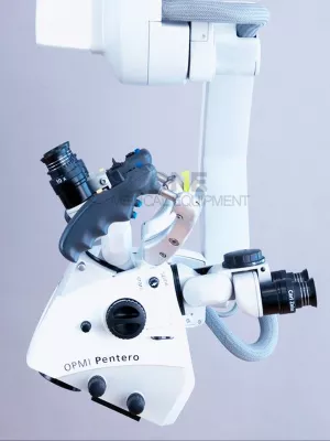
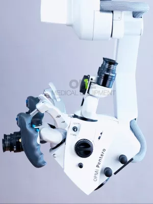
Carl Zeiss OPMI Pentero Surgical Microscopes
 First released in 2004, Zeiss OPMI Pentero surgical microscope has integrated intra-operative diagnostics and the full digital video chain into a user-friendly microscope design.
The Zeiss OPMI Pentero surgical microscope features apochromatic optics that deliver crystal-clear images, sharp details, and natural colors. The OPMI Pentero has 20% more light than previous models with spot illumination to precisely adjust the light cone. The Pentero has integrated high-speed autofocus that automatically delivers sharp images regardless of magnification. With the overhead design of this microscope, the suspensions system can be placed in any position, even behind the surgeon.
Features:
- Automated functions such as AutoBalance and AutoDrape
- Image-guided surgery with MultiVision data injection
- Integrated digital visualization, optionally with integrated high-definition (HD) camera head
- DICOM networking capabilities
- Touchscreen operation
Zeiss OPMI Pentero Specifications
Dimensions
- Height: 81.1” (206 cm)
- Width: 28.97” (73.6 cm)
- Depth: 28.97” (73.6 cm)
Magnification System
- Motorized zoom, apochromatic, 1:6 ratio
- Magnification displayed on the touchscreen and in the ocular (on demand)
- User-specific start position
Focusing System
- Varioskop, apochromatic, 200–500 mm working range
- Internal, motorized, continuous adjustment
- Magnification linked adjustment of focus speed
- High-speed laser autofocus, accurate to +/- 0.5 mm (Class II Laser)
- Visual focusing aid with two converging laser spots
- Working distance displayed on a touchscreen and in the ocular (on demand)
- User-specific start position
MultiVision System
- Integrated data display with shutter function
- SVGA 800 x 600, color, 50-60 Hz
- Color, binocular, injection and superimposition of contours and data
- Supported external data signals
- Computer data (VGA Signal)
- I.e. data from navigation systems
- Y/C video data (PAL / NTSC)
- I.e. data from endoscopy systems
- Computer data (VGA Signal)
- Superimposition of system information (focus, zoom, light)
- Injection of the touchscreen user interface into the eyepiece for sterile control of the system
Tubes and Co-Observation
- Main tube: 0–180° rotatable
- Eyepieces 10x/21B, 12.5x/18B
- Integrated beam splitter for lateral and face-to-face co-observation
- Stereo co-observation tube remains fixed when tilting the OPMI
- Spine adapter for symmetric face-to-face configurations
- Integrated rotary tube adapters
AutoDrape Systems
- Integrated vacuum system to remove air from sterile drape for fast and easy draping
Illumination System
- Superlux 330 light source with two 300 W Xenon daylight character lamps
- Integrated light source and light guide
- Integrated two-way illumination brightens shadows
- Variable spot illumination, minimum diameter 10 mm
- Semi-automatic lamp exchange
- Display of remaining lamp life on Touchscreen
- Brightness regulation via handgrips
- Magnification dependant automatic brightness adjustment
- Synchronized camera flash system
AutoBalance
- AutoBalance of the microscope, suspension system or entire system by pushing a button
- Microscope AutoBalance independent of position or accessories
Hospital Workflow Integration
- Varioskop, apochromatic, 200–500 mm working range
- Internal, motorized, continuous adjustment
- Magnification linked adjustment of focus speed
- High-speed laser autofocus, accurate to +/- 0.5 mm (Class II Laser)
- Visual focusing aid with two converging laser spots
- Working distance displayed on a touchscreen and in the ocular (on demand)
- User-specific start position
MultiVision System
- LAN interface and modem
- Microphone and speaker
- Patient data management allowing archival of image, video and audio data service file
- Remote service interface
Integrated Digital Video Chain
- 3CCD-Video camera PAL/NTSC:
- Video output on touchscreen
- Digital video outputs: Firewire/DV and Progressive Scan (VGA)
- Analog video outputs: FBAS (BNC), Y/C, RGB
- Stereo camera
- Image capture:
- Image freeze function
- Image capture as TIFF, JPG, BMP
- Image annotation
- Still image archiving via CD/DVD/USB and optional DICOM** interface
- Digital video recording system:
- MPEG2 recording
- Parallel HD/DVD recording
- Editing function
On Apr 14, 2012 Carl Zeiss Meditec, Inc., introduced the all-new PENTERO 900 surgical microscope system at the 79th Annual Meeting of the American Association of Neurological Surgeons (AANS) in Denver, Colo. Based on the breakthrough OPMI® Pentero surgical visualizationplatform launched in 2004, PENTERO 900 offers innovative new features and customer benefits such as:
HD quality for education, presentations and patient documentation
PENTERO 900 offers razor-sharp images in HD quality – camera, recorder, editor and touchscreen monitor are all contained in a fully integrated HD chain. The playback of high resolution HD images and videos is beneficial for surgeons during live demonstrations, resident education, presentations and patient documentation.
New fields of application for fluorescence
The YELLOW 560 option visualizes fluorescence in the wavelength range from 540 to 690nm supporting new fields of application and research. YELLOW 560 effectively complements the existing ZEISS fluorescence portfolio including BLUE 400, INFRARED 800 and FLOW 800 modules, paving the way for future application developments.
Comfortable working positions with new foldable binocular tube
The Foldable Tube f170/f260 combines brilliant apochromatic optics and a revolutionary design for maximum visualization flexibility, magnification potential and positioning ergonomics. It offers surgeons an unprecedented range of comfortable working positions, even at extreme surgical approach angles. The PROMAG™ function instantly yields a 50 percent gain in magnification for delicate and detailed procedures.
”When we introduced OPMI Pentero to the market in 2004, we set new, pioneering standards in the user-oriented enhancement of surgical microscope design. With the new features on PENTERO 900, we are propelling the microsurgical platform into a new dimension,” said Dr. Ludwin Monz, president and CEO of Carl Zeiss Meditec.