
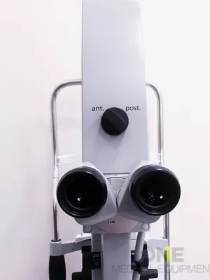
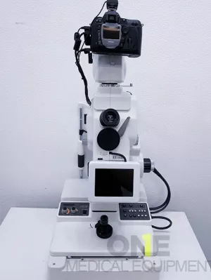
OCULUS Pentacam Topographer Classic for Sale – Patient-Ready Ophthalmic Diagnostic System which includes Basic Software, Refractive & Cataract Software, Wavefront Holladay Modules. Fully calibrated and patient-ready, a 6-month warranty, and a 14-day money-back guarantee for complete peace of mind.
The OCULUS Pentacam Classic Topographer is a fully patient-ready ophthalmic diagnostic system designed for precise corneal tomography and comprehensive anterior segment analysis. Ideal for ophthalmology clinics and eye care professionals, this advanced system delivers accurate and reliable diagnostic imaging.
Includes;
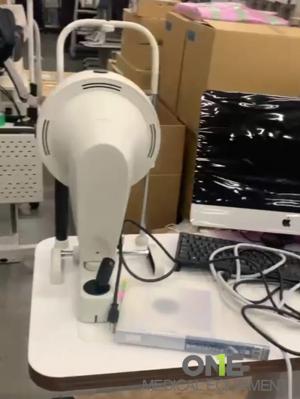
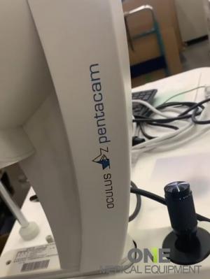
- OCULUS Pentacam Topographer main unit
- Basic Software plus Refractive & Cataract Software
- Wavefront, Holladay Modules
- Power and data cables
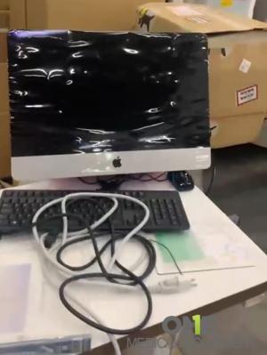
- Computer workstation
- Original software license
- Calibration and patient-ready certification
- User manual and training materials
The OCULUS Pentacam is a world-renowned corneal tomographer and topographer used by ophthalmologists and optometrists for comprehensive anterior segment analysis. The OCULUS Pentacam is a corneal tomographer and topographer, a specialized diagnostic device used in ophthalmology and optometry to analyze the front and back surfaces of the cornea (the clear, dome-shaped surface of the eye), as well as the anterior segment (front part of the eye including the anterior chamber, iris, and lens). The Pentacam uses a rotating Scheimpflug camera to capture high-resolution cross-sectional images of the anterior eye. It takes up to 50 slit images in less than 2 seconds, creating a 3D model of the cornea and anterior chamber.
Key Features
- Rotating Scheimpflug Camera Technology – Captures 3D cross-sectional images of the anterior eye in seconds.
- Full Corneal Tomography – Measures both anterior and posterior corneal surfaces for accurate curvature and thickness analysis.
- Pachymetry Mapping – Provides detailed corneal thickness distribution essential for refractive surgery planning and glaucoma assessment.
- Keratoconus Detection – Early detection with built-in indices to identify ectatic changes.
- Anterior Chamber Analysis – Measures chamber depth, volume, and angle for cataract and IOL planning.
- Lens Densitometry – Quantifies lens opacity for cataract evaluation.
- User-Friendly Software Interface – Easy navigation, automated reports, and export options for EMR integration.
Applications
- LASIK / PRK screening
- Cataract surgery planning
- Glaucoma assessment
- Keratoconus and ectasia monitoring
- Post-refractive surgery follow-up
- Anterior segment research
Brochure OCULUS Pentacam Topographer