
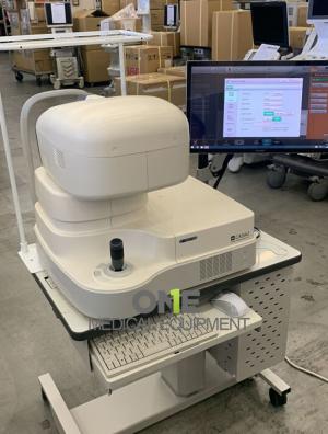
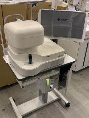
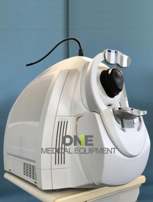
Sale Pre-Owned, Patient-Ready TOMEY Casia 2 OCT – Swept-Source Anterior Segment OCT High-speed, high-resolution anterior eye imaging for ophthalmology and optometry clinics.
Fully patient-ready Tomey CASIA 2 OCT has been professionally inspected, calibrated, and certified to meet manufacturer performance standards. It delivers high-resolution, cross-sectional, and 3-D images of the anterior eye structures – the cornea, anterior chamber, iris, and crystalline lens – with remarkable speed and accuracy. The Casia 2 empowers clinicians to diagnose and manage glaucoma, cataract, and corneal disease with confidence, at a fraction of the cost of a new device.
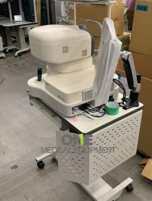
Package sale includes:
- Tomey CASIA 2 Anterior ocular segment 3D OCT
- Electric optical stand CT-700
- PCOS: Windows 8
TOMEY Casia 2 OCT
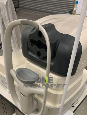
The TOMEY Casia 2 OCT is an advanced, non-contact imaging system that revolutionizes anterior eye diagnostics. With swept-source OCT technology, it delivers fast, deep, and highly detailed images of the cornea, iris, and lens — helping clinicians make confident diagnoses, plan surgeries with precision, and monitor outcomes with accuracy. Ideal for ophthalmology clinics, eye hospitals, and research institutions, the Casia 2 represents the future of anterior segment imaging. The Casia 2 combines speed, precision, and patient comfort in a single platform. Its non-contact, swept-source technology eliminates patient discomfort and reduces infection risk. The auto-alignment feature ensures quick scans with consistent accuracy, even for inexperienced operators.
Compared to older models or spectral-domain OCTs, the Casia 2 offers:
- Wider scan coverage for the entire anterior segment
- Improved repeatability and reproducibility
- Deeper visualization through opaque media (e.g., corneal scars or edema)
These capabilities make it one of the most advanced anterior segment OCTs available globally.
Key Features of TOMEY Casia 2 OCT
- High-speed swept-source technology (1310 nm wavelength, 50,000 A-scans/sec)
- 3D anterior segment imaging with angle analysis for glaucoma assessment
- Full corneal topography and pachymetry mapping for refractive surgery planning
- Automatic measurement of anterior chamber depth, lens position, and angle width
- Non-contact and patient-friendly design ensures comfortable imaging
- Comprehensive software for image analysis, IOL calculations, and progression monitoring
The TOMEY Casia 2 OCT is a state-of-the-art swept-source anterior segment Optical Coherence Tomography (OCT) system designed and manufactured by TOMEY Corporation, Japan, a world leader in ophthalmic diagnostic technology. This advanced imaging device provides high-resolution, cross-sectional, and three-dimensional images of the anterior segment of the eye, including the cornea, anterior chamber, iris, and crystalline lens. With its non-contact scanning technology and ultra-fast imaging speed, the Casia 2 delivers accurate, detailed images that are essential for the diagnosis and management of various eye conditions such as glaucoma, cataract, and corneal diseases.
Advanced Swept-Source OCT Technology
Unlike traditional time-domain or spectral-domain OCT systems, the Casia 2 uses Swept-Source OCT (SS-OCT) technology operating at a 1310 nm wavelength. This longer wavelength allows for:
- Deeper tissue penetration, enabling visualization of structures behind the iris and lens.
- Faster image acquisition, with speeds up to 50,000 A-scans per second, minimizing motion artifacts.
- High signal stability, even through opaque or cloudy corneas.
This makes the Casia 2 an invaluable tool for both clinical ophthalmology and research applications where speed, depth, and precision are critical.
Comprehensive Anterior Segment Analysis
The Casia 2 performs a complete analysis of the entire anterior segment in a single scan. It automatically measures:
- Corneal curvature and thickness (pachymetry)
- Anterior chamber depth and volume
- Iris and lens position
- Angle width and configuration
These parameters are vital for evaluating glaucoma risk, preoperative cataract planning, corneal refractive surgery, and postoperative outcomes.
3D Imaging and Angle Assessment
With the Casia 2, clinicians can generate three-dimensional reconstructions of the anterior segment, allowing detailed visualization of the iridocorneal angle, corneal layers, and lens structure.
This capability enables:
- Identification of narrow or closed angles in glaucoma patients.
- Visualization of the anterior chamber for IOL (Intraocular Lens) placement assessment.
- Comparison of pre- and post-surgical anterior eye configurations.
The 3D imaging module also supports cross-sectional slicing, giving ophthalmologists flexible tools for custom analysis.
Integrated Analysis Software
The TOMEY Casia 2 Anterior Segment Analysis Suite provides:
- Intuitive graphical user interface with auto-alignment and auto-scan capture.
- Measurement tools for corneal topography, pachymetry, and anterior chamber geometry.
- Comprehensive database for patient management and progression tracking.
- Export options in DICOM, PDF, and JPEG for EHR integration.
This seamless integration simplifies daily workflow in busy eye clinics and research institutions.
Clinical Applications
The TOMEY Casia 2 OCT is used extensively in:
- Glaucoma Diagnosis and Management
- Detect and classify open-angle or angle-closure glaucoma.
- Monitor structural changes over time.
- Cataract and Refractive Surgery Planning
- Measure corneal thickness, curvature, and anterior chamber depth.
- Evaluate IOL placement and lens position post-surgery.
- Corneal Disease Evaluation
- Assess keratoconus, corneal scars, and postoperative healing.
- Perform pachymetry mapping to detect ectasia or thinning.
- Anterior Segment Research
- Quantitative imaging for clinical studies and surgical innovation.
Technical Specifications
| Technology | Swept-Source OCT (1310 nm) |
|---|---|
| Scan Speed | 50,000 A-scans per second |
| Axial Resolution | 10 µm or better |
| Scan Depth | Up to 13 mm |
| Scan Width | Up to 16 mm |
| Display | 3D anterior segment visualization with angle analysis |
| Software | CASIA 2 Anterior Analysis Suite (Windows-compatible) |
| Manufacturer | TOMEY Corporation, Japan |