
Zeiss Cirrus 500 HD-OCT for Sale, Patient-Ready Optical Coherence Tomography System, has been fully refurbished by Zeiss certified Technicians and comes with a 12-month parts and service warranty.
This refurbished Zeiss Cirrus 500 HD-OCT offers the same reliability and precision as a new system, at a fraction of the cost. Each device is fully serviced, calibrated, and quality checked to meet clinical performance standards ready to use the moment it arrives at your practice. Every refurbished Zeiss Cirrus 500 OCT undergoes a strict multi-point inspection by certified biomedical engineers.
We ensure:
- 100% functional verification of all hardware and optics
- Calibration to manufacturer standards
- Software reinstallation and performance optimization
- Replacement of worn parts with OEM components
- Thorough cleaning and cosmetic restoration
- Final image quality and safety testing
Included
- Refurbished Zeiss Cirrus 500 HD-OCT
- Computer with pre-installed Zeiss software
- LCD monitor, keyboard, and mouse
- Power cables and accessories
- User manual
- 6 month warranty and technical support
- 30 days money back guarantee
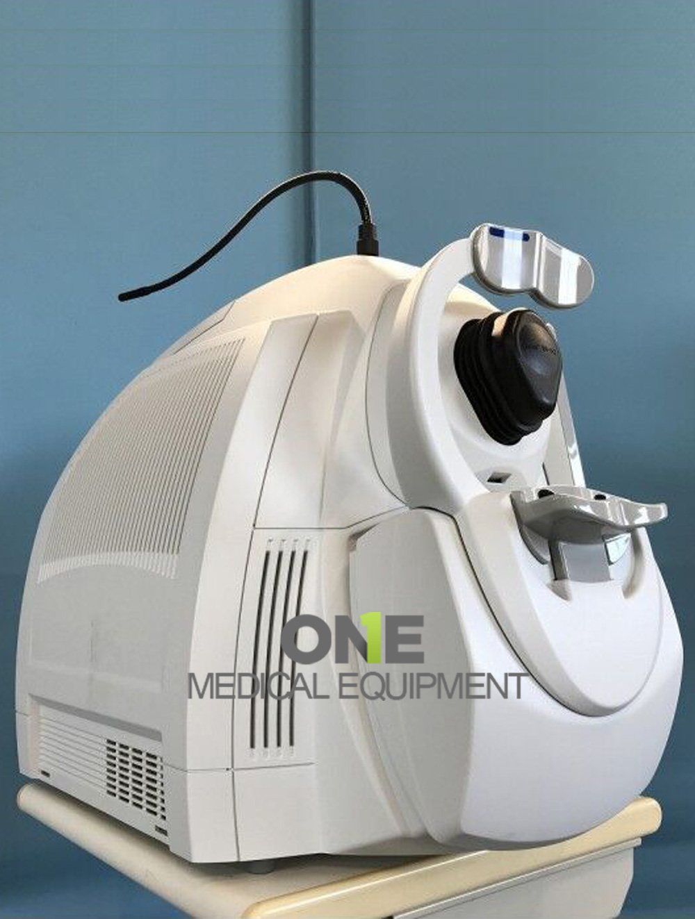
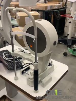
Zeiss Cirrus 500 HD-OCT – High-Definition Optical Coherence Tomography System
The Zeiss Cirrus 500 HD-OCT is a high-performance Optical Coherence Tomography (OCT) system engineered for retinal, macular, and optic-nerve head imaging. Developed by Carl Zeiss Meditec, a global leader in ophthalmic diagnostics, the Cirrus 500 delivers exceptional image clarity, reproducibility, and workflow efficiency empowering eye-care professionals to detect, diagnose, and monitor ocular diseases with precision and confidence.
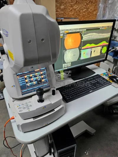
Designed for clinical reliability and ease of use, the Cirrus 500 integrates advanced scanning optics, sophisticated analysis software, and a compact footprint ideal for ophthalmology clinics, hospitals, and optometry practices.
The Zeiss Cirrus 500 HD-OCT is one of the most widely used OCT systems in ophthalmology and optometry. It delivers clear, detailed retinal images for accurate diagnosis and patient monitoring.
The Zeiss Cirrus 500 HD-OCT is a trusted imaging system designed for high-definition retinal and optic nerve analysis.
Key Features & Benefits
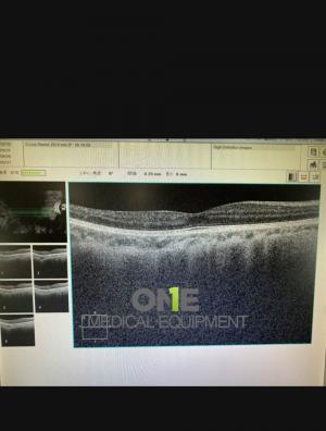
High-Definition Imaging
- Captures ultra-high-resolution retinal cross-sections with axial resolution around 5 microns, enabling detailed visualization of retinal layers.
- Provides both qualitative and quantitative analysis for accurate diagnosis of retinal pathologies.
Fast Scanning Performance
- Scan speed of 27,000 A-scans per second reduces motion artifacts and enhances patient comfort.
- Multiple scan patterns (Macula Cube 512×128, Optic Disc Cube 200×200, Radial Line, Raster Line) deliver comprehensive coverage in seconds.
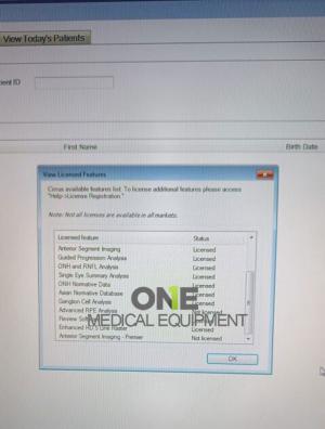
Advanced Analysis Software
- Automated segmentation of retinal layers for precise thickness maps.
- Glaucoma and macular progression analysis modules with normative databases.
- DICOM-compatible for seamless integration with EMR systems.
- Intuitive user interface simplifies exam setup, image review, and report generation.
IIntegrated Data Management
- Built-in Cirrus Review Software allows clinicians to view and analyze patient data anywhere in the clinic.
- Long-term data storage supports trend analysis for disease progression and treatment outcomes.
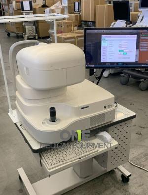
Compact & Ergonomic Design
- All-in-one tabletop configuration with minimal footprint.
- Operator-friendly joystick and patient alignment system ensure quick, comfortable examinations.
Reliable Zeiss Quality
- Engineered with durable components and precision optics.
- Known for longevity, minimal maintenance, and consistent imaging performance.
The Zeiss Cirrus 500 HD-OCT supports a wide range of ophthalmic diagnostic applications:
- Glaucoma detection and monitoring (optic-nerve and RNFL analysis)
- Macular degeneration evaluation (AMD, CNV, drusen)
- Diabetic retinopathy and macular edema assessment
- Retinal-layer mapping and vitreoretinal interface analysis
- Pre-and post-surgical evaluation of retinal and optic-nerve conditions
OCT Imaging:
- Methodology: Spectral domain OCT
- Optical source: Superluminescent diode (SLD), 840 nm
- Scan speed: 27K- 68K A-scans per second
- A-scan: 2.0 mm (in tissue), 1024
- Axial resolution: 5 μm (in tissue)
- Transverse resolution: 15 μm (in tissue)
Fundus Imaging:
- Methodology: Love OCT Fundus
- Live fundus image: During alignment
- Optical source: Superluminescent diode (SLD), 840 nm
- Field of view: 36 degrees W x 30 degrees H
- Frame rate: > 1.7 Hz
- Transverse resolution: 45 μm (in tissue)
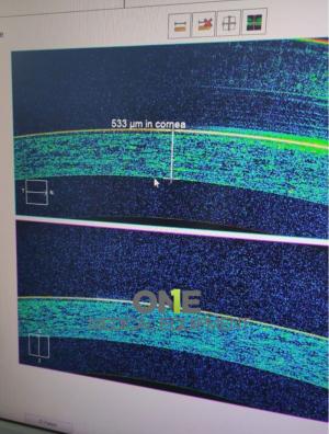
Iris Imaging:
- Methodology: CCD camera
- Resolution: 1280 x 1024
- Live iris image: During alignment
Electrical and Physical:
- Weight: 76 lbs (34 kg)
- Dimensions of instrument: 26L x 18W x 21H (in), 65L x 46W x 53H (cm)
- Dimensions of table: 39L x 22W (in), 99L x 56W (cm)
- Fixation: Internal, external
- Internal fixation focus adjustment: -20D to +20D (diopters)
- Electrical rating (115V): Single Phase, 100–120V~ systems: 50/60Hz, 5A
- Electrical rating (230V): Single Phase, 220–240V~ systems: 50/60Hz, 2.5A
Internal Computer:
- Operating system/processor: Windows 7, 4th generation i7 Intel processor
- Memory: 16 GB
- Hard drive/internal storage: ≥ 2 T (> 200,000 scans)
- Display: Integrated 19“ color flat panel display
- USB ports: 6 ports
| Parameter | Specification |
|---|---|
| Technology | Spectral-domain OCT |
| Axial Resolution | ~5 μm |
| Transverse Resolution | ~15–20 μm |
| Scan Speed | 27,000 A-scans/second |
| Scan Depth | 2.0 mm in tissue |
| Scan Patterns | Macula Cube 512×128, Optic Disc Cube 200×200, Line Raster, Radial, 3D Cube |
| Field of View | 6 mm × 6 mm |
| Display | Integrated LCD monitor |
| Connectivity | USB / DICOM / Network |
| Power | 100–240 V AC, 50–60 Hz |
| Dimensions | Approx. 19 in (W) × 18 in (D) × 16 in (H) |
| Weight | ~50 lbs (23 kg) |