
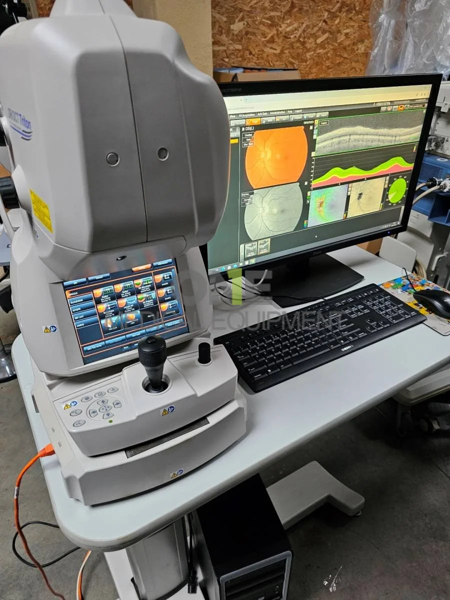
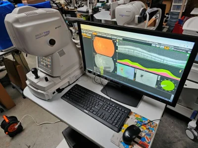
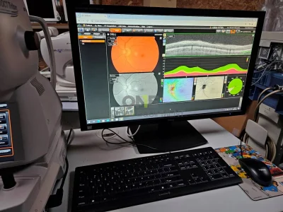
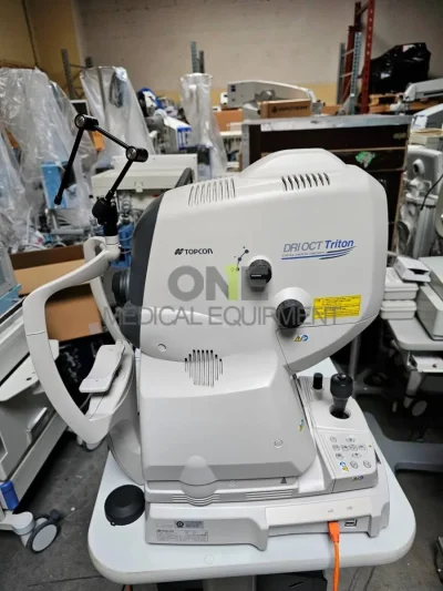
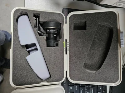
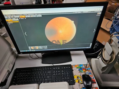
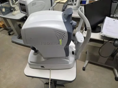
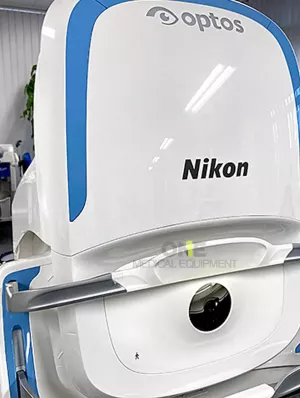
Certified pre-owned Topcon DRI Triton Plus OCT Anterior for sale, excellent patient ready condition with PC set and Anterior eye lens. 1050nm OCT for Posterior and anterior segment OCT and optional OCT Angiography imaging. Color, red free, FA and FAF photography.
All items are sold in as-is condition with 6 month warranty.
Includes everything and ready to work:
- Topcon DRI OCT Triton Plus
- PC set with rack
- Electric optical stand
- Printer
- Anterior eye lens unit AA-1
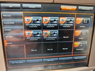
- ** OCT Angiography can be performed in combination with IMAGEnet 6.
Topcon DRI Triton Plus OCT
The DRI OCT Triton combines the world’s first Swept Source OCT technology with multimodal fundus imaging. Triton uses Swept-Source technology to allow visualization into the deepest layers of the eye – even through cataracts, hemorrhages, gas bubbles, and other media opacities, making it possible for more patients to be imaged.
The fast 100 kHz scanning speed and invisible scan beam rapidly capture detailed images, resulting in fewer motion artifacts and stunning image quality. Decrease chair time and improve your clinical workflow with fewer rescans and multimodal imaging including, SS-OCT, True-Color, Digital Red-free, and optional Anterior Segment OCT.
Triton (plus) adds fluorescein angiography (FA) and fundus autofluorescence (FAF) to the Triton.
Key features
- Swept-Source OCT with 100,000 A-scan/sec scanning speed
- 1050nm wavelength
- Multimodal imaging: True Colour Fundus Photography, OCT-Angiography(1), Wide Field for OCT and OCT-A(1), Anterior Segment(1), FA*, FAF*
Image Quality
Triton’s Swept Source OCT with its 100kHz scanning speed and 1,050nm wavelength results in a clear and detailed images even for the deepest layers of the eye with short acquisition time. Visualise not only the retina and vitreous, but also the choroid and sclera2.
NEW: Discover Smart Denoise1, Topcon's unique AI algorithm which reduces artifacts and increases contrast on OCT and OCT-A1.
Diagnostic capability
Seeing deeper makes it possible to have a better understanding of many ocular pathologies2. With features such as OCT Angiography1, Fundus Autofluorescence* and En Face OCT, Triton empowers clinicians with multimodal imaging capability to help assess and preserve patient’s eye health.
Practice efficiency
The Triton’s automated functions, including single scan capture and SMARTTrackTM system, are designed to optimise your practice workflow by simplifying data capture, analysis and follow-up.
Swept Source OCT Imaging
1,050nm wavelength
The longer wavelength light provides better tissue penetration, allowing visualisation into the deepest layers of the eye2.
Swept Source OCT technology and scanning speed of 100,000 A-scans/sec
The fast scanning speed of 100,000 A-scans/sec enables capture of clear B-scans3 by acquiring more A-scans within a given image acquisition time. This helps to reduce artifacts from involuntary eye movements such as saccades and blinks.
Invisible scan lines
The invisible 1,050nm wavelength light helps patients concentrate on the fixation target during the scan, reducing involuntary eye movement. It supports more efficient workflow in a practice by reducing the need to rescan.
Swept Source OCT incorporates multimodal fundus imaging
True Colour6 Fundus images
The DRI OCT Triton offers a true colour, non-mydriatic fundus image. Fluorescein Angiography (FA)* and Fundus Autofluorescence (FAF)* are available to enhance the diagnostic capability of Triton Plus.
The all-in-one device supports efficient workflow in practice. Swept Source OCT incorporates multimodal fundus imaging. DRI OCT Triton can acquire the OCT and fundus image in a single capture.
PinPoint™ Registration identifies the location of the B-scan on the fundus image. Comparison between the B-scan and fundus image can support clinical efficiency during diagnosis.
NEW: Wide Field OCT and OCT-A
The optional Wide-Field attachement lens enables the capture of scans up to 21mm in length. Gather more clinical insights with OCT and OCT-A visualisation. Wide-Field imaging is valuable in a wide variety of conditions.
NEW: Anterior Segment Metrics
Triton’s optional anterior segment imaging capabilities allow for visualisation of the cornea, anterior chamber angle, iris and anterior sclera and integrates quantitative analysis.
The new anterior segment feature reaffirms Triton’s value in eye care settings with detailed reports
Follow-up Function
This function allows you to retrieve and re-analyse the same location at follow-up, for comparison of past and current images. All an operator needs to do is simply select the past data and Triton automatically captures the same area.
SS-OCT Angiography
Topcon’s SS-OCT Angio combines OCT angiography with Swept-Source technology.
OCTARA™, a proprietary image processing algorithm, provides highly sensitive angiographic detection4 allowing for visualisation of vascular structures even in the choroid and deeper retinal layers.
OCTARATM
OCTARATM is the image processing technology which extracts the signal changes derived from vascular flow using multiple OCT B-Scans acquired at the same position. It demonstrates high sensitivity for the detection
of low blood flow in microvasculature4.
High-sensitivity Imaging and Deeper Intravascular Flow Visualisation
Swept Source technology and OCTARA™ allow the deeper structures to be visualised with less depth dependent signal roll-off3. Additionally, the 1μm wavelength makes OCT Angiography imaging possible for patients with media opacities.
Rapid Scanning, Real Time Eye Tracking
At 100,000 A-Scans per second coupled with invisible5 scanning lines and the SMARTTrack™ eye tracking system, Triton quickly captures a dense data set and provides an En Face OCT Angiography image of the retinal microvascular flow network4.
Technical Specifications
| Observation & Photography of Fundus Image | Photography Type: Color, FA*, FAF*, Red-free** |
| Picture Angle: 45° Equivalent 30° (Digital Zoom ) |
|
| Operating Distance: 34.8mm | |
| Photographable Diameter of Pupil: Normal:φ4.0mm or more Small pupil diameter: φ3.3mm or more |
|
| Observation & Photography of Fundus Tomogram | Scanning Range (on fundus): Horizontal Within 3 to 12mm Vertical Within 3 to 12mm |
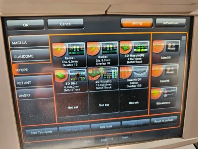
| Scan Pattern: 3D scan Linear scan (Line-scan/Cross-scan/Radial-scan) |
|
| Scan Speed: 100,000 A-Scans per second | |
| Lateral Resolution: 20μm | |
| In-depth Resolution: Digital: 2.6μm Optical function: 8μm |
|
| Photographable Diameter of Pupil: φ2.5mm or more | |
| Observation & Photography of Fundus Image / Fundus Tomogram | Fixation target; Internal fixation target : Dot matrix type organic EL The display position can be changed and adjusted. The displaying method can be changed. Peripheral fixation target : This is displayed according to the internal fixation target displayed position. External fixation target |
| Observation & photography of anterior segment*** | Photography type: IR |
| Operating distance: 17mm | |
| Observation & photography of anterior segment tomogram*** | Operating distance: 17mm |
| Scan range (on cornea): Horizontal Within 3 to 16mm Vertical Within 3 to 16mm |
|
| Scan pattern: 3D scan Linear scan (Line-scan/Radial-scan) |
|
| Scan speed: 100,000 A-Scans per second | |
| Fixation target: Internal fixation target External fixation target |
|
| Electric Rating | Power Source: Voltage: 100-240V Frequency: 50-60Hz |
| Power input: 250VA | |
| Dimensions / Weight | Dimensions: 320-359 mm(W) X 523-554 mm(D) X 560-590 mm(H) |
| Weight: 21.8kg (DRI OCT Triton) 23.8kg(DRI OCT Triton plus) |