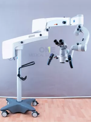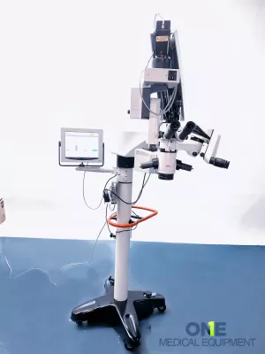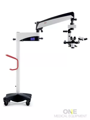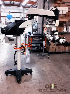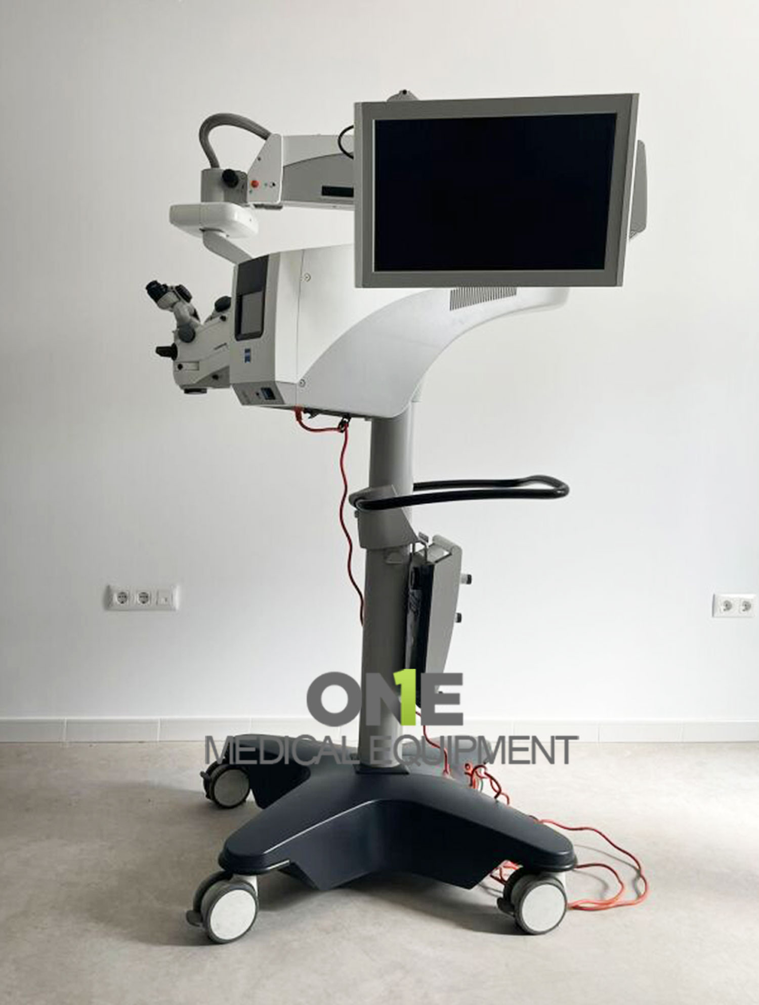
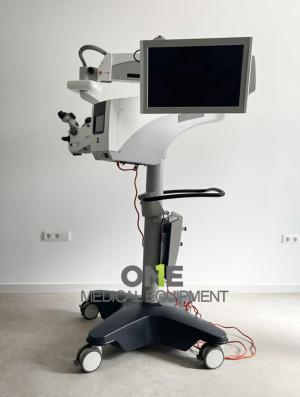
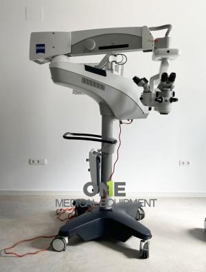
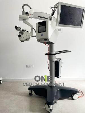
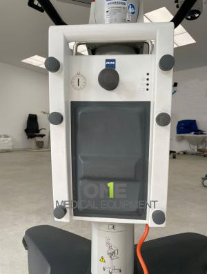
ZEISS Opmi Lumera 700 Surgical Microscopes for sale, Fully Serviced and Certified, Premium Patient, Ready Ophthalmic Microscope. Sold as is with 12-month warranty and 30 days money back guarantee.
Specifications:
- Zoom System: Motorized, apochromatic optics, 1:6 zoom ratio
- Magnification: 0.4× – 2.4×
- Focus: Motorized/electric, 70 mm range
- Objective Lens: Standard f = 200 mm (optional: f = 175 mm or f = 225 mm with support ring)
- Binocular Tube: Invertertube E (alternatives: Invertertube, 180° swivel tube f = 170 mm, inclined tube f = 170 mm)
- Eyepieces: 10× wide-angle (optional 12.5×)
- SCI Lighting: Coaxial and full-field
- Fiber-Optic (Superlux Eye)
- Xenon short-arc lamp with HaMode filter
- Manual backup lamp integrated into housing
- LED Fiber-Optic: Daylight-like color temperature, Approx. 50,000 hours lifespan at 50% intensity, HaMode filter + 25% gray filter
- Universal Filters: Blue-blocking filter, Optional fluorescence filter
- Integrated Slit Illumination: Widths: 0.2 / 2 / 3 / 4 mm, Height: 12 mm
Included;
- ZEISS Opmi Lumera 700 Surgical Microscopes
- Main microscope body & illumination module
- Footswitch control unit
- Assistant binocular (optional)
- Power supply & cables
- Warranty certificate
- User manual & calibration report
If you’re looking for a high-performance surgical microscope trusted by ophthalmic surgeons worldwide, the ZEISS Opmi Lumera 700 remains one of the most advanced and reliable systems available. Our refurbished patient-ready ZEISS Lumera 700 units are fully inspected, recalibrated, and certified by biomedical engineers — delivering as-new performance at a fraction of the cost.
ZEISS OPMI LUMERA 700 Seeing to succeed with the microscope for every ophthalmic specialty
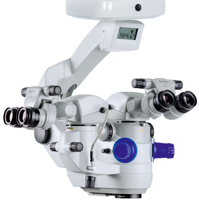 Whether preserving or restoring a patient’s sight, the OPMI LUMERA 700 from ZEISS is the surgical microscope for every ophthalmic specialty. Experience markerless IOL alignment and integrated intraoperative OCT3 imaging – all in one device – from the ophthalmic microscope market leader.
ZEISS OPMI LUMERA 700 is part of our commitment to helping you succeed in your OR. It’s also part of the ZEISS Cataract Suite, which includes leading products designed to work together for markerless toric IOL alignment.
- Markerless toric IOL alignment
- Improved visualization
- Teaching possibilities
One example is with the OPMI LUMERA 700 from ZEISS, an
operating microscope ideally suited for every ophthalmic
surgery speciality. Experience markerless IOL alignment and
integrated intraoperative OCT* imaging – all in one device.
Seeing to succeed in cataract surgery
Precise and efficient markerless toric IOL alignment. With ZEISS CALLISTO eye markerless alignment, manual marking steps can be skipped altogether for an efficient and precise toric IOL alignment to reduce residual astigmatism
For cataract surgeries, ZEISS OPMI LUMERA 700, with its well-known patented SCI illumination, ZEISS optics and CALLISTO eye from ZEISS provides the best anterior views and precise assistance functions.
Cataract assistance functions for every step of the surgery
The assistance functions of ZEISS CALLISTO eye are completely surgeon-controlled – with either the foot control panel or handgrips.
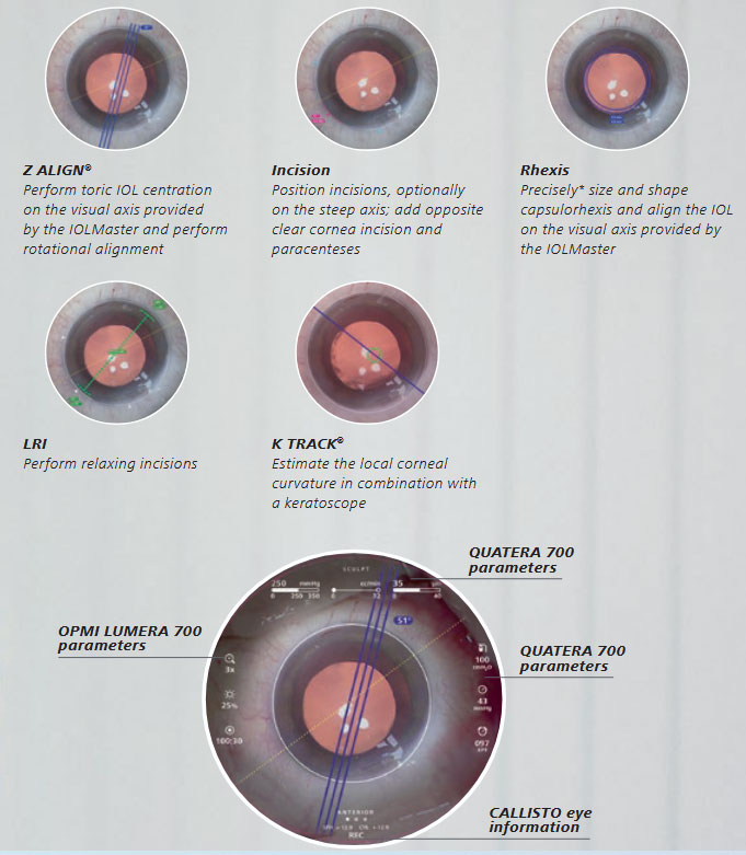
Efficient markerless IOL alignment Starting with a biometry reference image from the IOLMaster from ZEISS, data is transferred smoothly to CALLISTO eye. This data is used to create overlays in the eyepiece. Save time, increase efficiency and reduce residual astigmatism when you:
- skip manual preoperative marking
- skip manual data transfer
- skip manual intraoperative marking
Efficient surgery setup
The image quality check supports you to optimize light intensity, magnification and centration of the microscope toefficiently set up the reference axis. The well-proven* eye tracking automatically compensates for eye movements and supports the use of the assistance functions.
Seeing to succeed in glaucoma surgery, Improved visualization
As minimally invasive glaucoma surgery (MIGS) and canaloplasty procedures evolve, intraoperative OCT plays an increasingly important role, particularly for monitoring implants such as stents in difficult to see spaces. The integrated intraoperative OCT* images of the ZEISS OPMI LUMERA 700 enable a clear visualization of the device placement to help achieve excellent outcomes
More information to support your decisions during surgery
Integrated intraoperative OCT* visualizes the orientation and placement of the MIGS implant, supporting surgical decisions and providing more information on outcomes. Distortion-free, computer enhanced intraoperative OCT* images visualize detailed structures in the natural physiological shape.
Stay focused on the area of interest
Save time by maintaining the selected intraoperative OCT* scan location with the new automatic XY tracker. In addition to the proven Z tracker, the XY tracker compensates for movements of the eye or the microscope
Protect the retina
Shield the retina from excessive light exposure with the integrated retina protection filter.
Flexible perspective for a better view
Tilt the microscope head as needed to better observe the iridocorneal angle
Seeing to succeed in cornea surgery, Reduce graft manipulation
Clinical results indicate that using intraoperative OCT* can reduce cell loss.** Studies show that intraoperative OCT* from ZEISS can lead to quicker decisions***, resulting in reduced manipulation time and, therefore less cell loss. The integrated intraoperative OCT* of the ZEISS OPMI LUMERA 700 visualizes the actual physiological shape of the cornea in two different scan views. Switch between views with a touch of the finger or tap of the foot to make your decisions faster.
Make faster decisions with two scan depths and a realistic view.
Quickly change between high-resolution OCT scans (2.9 mm scan depth in tissue) and large overview images (5.8 mm scan depth in tissue) to visualize and assess
graft orientation. Observe the natural physiological shape of the cornea with distortion-free intraoperative OCT* images. See how intuitive OCT image navigation is during surgery.
DMEK: save time with easy graft monitoring
Monitor the graft orientation and assess the interface with the patient´s cornea. Verify proper graft positioning as well as visualize fluid interface and graft adherence.
DALK: secure big-bubble procedure
OCT* imaging helps the surgeon during DALK to assess the dissection depth in order to reduce perforation risk and potentially improve the reproducibility of the big-bubble procedure
OPMI LUMERA 700 from ZEISS TECHNICAL DATA
| ZEISS OPMI LUMERA 700 | |
| Surgical microscope | Motorized zoom system with apochromatic lens, zoom ratio 1:6 |
| Magnification factor = 0.4 x – 2.4 x | |
| Focusing: electric / motorized, focus range: 70 mm | |
| Objective lens: f = 200 mm (optionally also f = 175 mm or f = 225 mm with support ring | |
| Binocular tube: Invertertube E (optionally also Invertertube, 180° swivel tube, f = 170 mm, inclined tube, f = 170 mm | |
| Wide-angle eyepiece 10 x (optionally also 12.5 x) | |
| Light source | SCI: Coaxial and full-field illumination |
| Fiber-optic illumination Superlux ® Eye: - Xenon short arc reflector lamp with HaMode filter - Backup lamp in lamp housing, can be slid into position manually |
|
| LED fiber-optic illumination: - Near-daylight color temperature - 50,000 hour lifetime at 50% light intensity - HaMode filter - 25% gray filter |
|
| For all light sources: - Blue blocking filter - Optional: Fluorescence filter |
|
| Integrated slit illuminator | Slit widths: 0.2 mm, 2 mm, 3 mm, 4 mm Slit height: 12 mm |
| XY coupling | Travel range: max. 61 mm x 61 mm Automatic centering at the touch of a button |
| Video monitor | 23.6” LCD display Resolution: 1,928 x 1,080 |
| Stand | Maximum permissible weight load of the spring arm: When the surgical microscope is attached to the arm (without tube, eyepiece or objective lens) and the XY coupling is also attached, a maximum of 9 kg of additional accessories can be attached to the spring arm |
| ZEISS intraoperative OCT | |
| OCT engine | SD (spectral domain) OCT Wavelength 840 nm Scanning speed 27,000 A-scans per second |
| Scan parameters | A-scan depth: 2.9 and 5.8 mm in tissue Axial resolution: 5.5 μm in tissue Scan length adjustable 3–16 mm Scan rotation adjustable 360° Scan modes for live and capture acquisition Live: 1-line, 5-lines, cross hair Capture: 1-line, 5-lines, cube |
| ZEISS RESIGHT family | |
| Mechanical data | Focus range with LH175 lens holder: 31 mm (position of intermediate image) |
| Focus range with LH200 lens holder: 38 mm (position of intermediate image) | |
| Rotation angle of lens revolver and holder: 0°–360° | |
| Lenses included | 60D, 128D |
| Weight | ZEISS RESIGHT 500 (manual): 0.45 kg ZEISS RESIGHT 700 (motorized): 0.50 kg |
| ZEISS CALLISTO eye panel PC | |
| Touch screen | Projected Capacitive Touch (PCT) with anti-reflective coating, scratch-proof |
| Processor | Intel ® Core i5 6442EQ 1.9 GHz |
| Hard drive | SSD for operating system, SATA HDD 1 TB for data |
| Display | Integrated 24” color flat screen with high luminosity and wide viewing angle |
| Video signals | PAL 576i50; NTSC 480i60; 1080i50; 1080i60 Only possible with camera models from Carl Zeiss Meditec AG |
| Ports | 1 × CAN-Bus, 2 × 1 Gigabit Ethernet, 5 × USB 3.0, 1 × potential equalization |
| Video input | 1 × Y/C, 1 × HD-SDI |
| Video output | 2 × HDMI |
| Connectivity | Integrated RJ45 10/100Base-T Ethernet port for connection to ZEISS OPMI LUMERA 700 and hospital network |
| Weight | ca. 10 kg |
| ZEISS CALLISTO eye software | |
| Version | 3.7 |